


Mr Stephen Price
Position: Clinical Consultant
Personal home page:
Email:
info@brain.cancer.cam.ac.uk
PubMed journal articles - click here
Mr Stephen Price is pleased to consider applications from prospective PhD students.
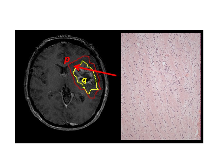
My research group is interested in using advanced MRI and PET imaging to study the area around brain tumours with a view to developing improved methods of local control of gliomas. In this 'peritumoural area' is tumour invasion into areas of normal, functioning brain. My work has two main themes i) using imaging to identify occult tumour invasion and predict the site where tumours will progress. This is currently being explored as part of the multi centre CRUK funded PRaM-GBM study that I am leading. The aim would be to aid planning of both radiotherapy and surgery. ii) Understanding the effect of treatments on the local surrounding brain. Most of this is to understand the effect of surgery on the surrounding brain to understand what structures need avoiding to prevent neurological deficits. I am also interested in the effect of radiotherapy on normal brain and am leading a national initiative to study this. This work involves both imaging white matter (DTI) and brain networks using resting state fMRI. We are particularly interested in visual and cognitive deficits and the impact these have on quality of life.
My other interest developed as part of a NIHR CLAHRC Fellowship involves the barriers with getting research into clinical practice and the impact that has allowing innovation lead improved patient benefit.
MRI Imaging; Image guided biopsies; Surgical targets; Translating research into clinical practice;
Symplectic Elements feed provided by Research Information, University of Cambridge
1. SJ Price, K Allinson, H Liu, NR Boonzaier, J-L Yan, VC Lupson and TJ Larkin (2016) IDH-1 mutated glioblastomas have a less invasive phenotype than IDH-1 wild type glioblastomas: a diffusion tensor imaging study. Radiology (in press).
2. J-L Yan, A van der Hoorn, TJ Larkin, NR Boonzaier, T Matys, SJ Price (2016) Extent of resection of peritumoural DTI abnormality as a predictor of survival in adult glioblastoma patients. Journal of Neurosurgery 2016 Apr 8:1-8. [Epub ahead of print].
3. SJ Price, Young AM, Scotton WJ, J Ching, LA Mohsen, NR Boonzaier, VC Lupson, JR Griffiths, MA McLean and TJ Larkin (2016) Multimodal MRI can identify perfusion and metabolic changes in the invasive margin of glioblastomas. Journal of magnetic resonance imaging 43(2): 487–494.