Researchers and medical experts from nine world-leading medical imaging centres across the UK come together to form an integrated infrastructure for standardising and validating cancer imaging biomarkers for clinical use.
The centres include the University of Cambridge, University College London, University of Manchester, University of Oxford, King’s College London, The Institute of Cancer Research, London and The Royal Marsden NHS Foundation Trust, Imperial College London, Newcastle University and University of Glasgow.
This unique UK infrastructure provides clinical researchers across the UK with open access to world-class clinical imaging facilities and expertise, as well a repository data management service, artificial intelligence (AI) tools and ongoing training opportunities.
The NCITA consortium, through engagement with NHS Trusts, pharmaceutical companies, medical imaging and nuclear medicine companies as well as funding bodies and patient groups, aims to develop a robust and sustainable imaging biomarker certification process, to revolutionise the speed and accuracy of cancer diagnosis, tumour classification and patient response to treatment.
Professor Evis Sala, Professor of Oncological Imaging in the Department of Radiology at the University of Cambridge, who co-leads our Advanced Cancer Imaging Programme said: "We are delighted to be part of NCITA and are coordinating a renal cancer imaging study for the network using a novel agent that is being developed in Cambridge.
"We are also establishing a shared image repository which will provide the large volume of data needed to 'train' new AI tools to accurately analyse patient scans in the future."
The NCITA initiative is funded by Cancer Research UK and will receive up to £10 million over 5 years.
The NCITA network is led by Prof Shonit Punwani, Prof James O’Connor, Prof Eric Aboagye, Prof Geoff Higgins, Prof Evis Sala, Prof Dow Mu Koh, Prof Tony Ng, Prof Hing Leung and Prof Ruth Plummer with up to 49 co-investigators supporting the NCITA initiative.
NCITA is keen to expand and bring in new academic and industrial partnerships as it develops.
Image caption and reference:
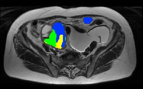
Phenotypically distinct areas (imaging habitats) of ovarian cancer can be identified by advanced analysis of imaging features derived from magnetic resonance imaging (MRI) and positron emission tomography (PET)/computed tomography (CT). These distinct imaging habitats (labeled blue, yellow and green), harbour distinct growth patterns with differential expression of hypoxia- and neovascularization-related markers and distinct somatic genetic alterations.
Weigelt B, Vargas HA, Selenica P, Geyer FC, Mazaheri Y, Blecua P, Conlon N, Hoang LN, Jungbluth AA, Snyder A, Ng CKY, Papanastasiou AD, Sosa RE, Soslow RA, Chi DS, Gardner GJ, Shen R, Reis-Filho JS, Sala E. Radiogenomics Analysis of Intratumor Heterogeneity in a Patient with High-Grade Serous Ovarian Cancer
JCO Precision Oncology June 2019; 3:1-9


