

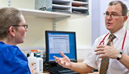
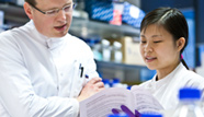
The first cancer patient in Europe has been scanned with a revolutionary imaging technique that could enable doctors to see whether a drug is working within a day or two of starting treatment.
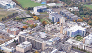
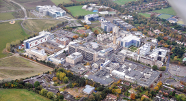
The patient is the first to take part in a new metabolic imaging trial of patients across a wide range of cancer types to be carried out by scientists at Addenbrooke’s Hospital. The study, which is funded by a Wellcome Trust Strategic Award, could show whether patients can stop taking drugs that aren’t working for them, try different ones and receive the best treatment for their cancer as quickly as possible.
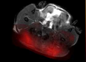
The rapid scan will allow doctors to map out molecular changes in patients, opening up potential new ways to detect cancer and monitor the effects of treatment.
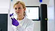
The hyperpolariser facility at Addenbrooke’s uses a revolutionary imaging technique called nuclear spin hyperpolarisation on a breakdown product of glucose called pyruvate. The pyruvate is labelled with a non-radioactive form of carbon called carbon 13 (C-13) which makes it 10,000 times more likely to be detected in a magnetic resonance imaging (MRI) scan.
Pyruvate is injected into the patient and tracked as the molecule moves around the body and enters cells. The scan monitors how quickly cancer cells break pyruvate down – a measure of how active the cells are that tells doctors whether or not a drug has been effective at killing them.
Professor Kevin Brindle, co-lead based at the Cancer Research UK Cambridge Institute, said: “We’re very excited to be the first group outside North America, and the third group world-wide, to test this with patients and we hope that it will soon help improve treatment by putting to an end patients being given treatments that aren’t working for them. Each person’s cancer is different and this technique could help us tailor a patient’s treatment more quickly than before.”
Dr Ferdia Gallagher, co-lead also funded by Cancer Research UK and based at the Department of Radiology at the University of Cambridge, said: “It’s fantastic that we can now try this technique in patients. We hope this will progress the way cancer treatment is given and make therapy more effective for patients in the future. This new technique could potentially mean that doctors will find out much more quickly if a treatment is working for their patient instead of waiting to see if a tumour shrinks.”
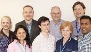
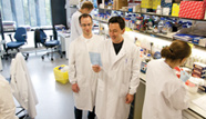
The joint project has involved a large number of collaborators in the University of Cambridge and at Addenbrooke’s hospital including Professor David Lomas (Radiology), Professor Ian Wilkinson (Department of Medicine), Dr Bristi Basu (Oncology) and Mrs Anita Chhabra (Pharmacy).
The facility and clinical trials are funded and supported by the Wellcome Trust, Cancer Research UK, Addenbrooke’s Charitable Trust, the Cambridge Cancer Centre, Cambridge University Hospitals NHS Foundation Trust, GE Healthcare and the University of Cambridge.