


Dr Adrian Ionescu
Position: Research Associate
Personal home page:
PubMed journal articles - click here
Dr Adrian Ionescu is pleased to consider applications from prospective PhD students.
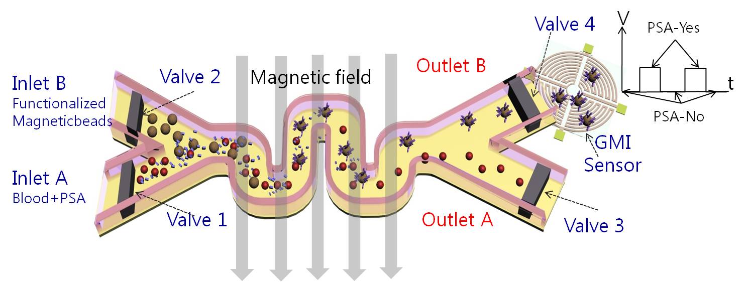
I currently work as a post-doctoral research associate at the Cavendish Laboratory, Department of Physics, University of Cambridge, in the Thin Film Magnetism (TFM) group with Prof. Crispin Barnes on an ERC International Training Network project, SIMDALEE2, studying low energy electron interactions with matter. In this position I have had the opportunity to work on a variety of contemporary experimental condensed matter physics research including spintronics, surface science, magnetism at the nanoscale and in low dimensional systems, spin polarised oxide interfaces, topological insulators, biomagnetism, magnetic biosensors, magnetic domain wall motion and magnetic storage technology. Over the last 10 years I have built up a strong track record with international recognition in these research areas, with 50 publications and two books to date. With some of these publications I have helped shape the new interdisciplinary field of Biomagnetics in which spintronic devices are used as bio-assays in life science applications.
Symplectic Elements feed provided by Research Information, University of Cambridge
Books:
1) Ionescu, A, Llandro, J and Darton, NJ (Editors).
“MAGNETIC NANOPARTICLES IN BIOSENSING AND MEDICINE”, Cambridge
University Press (and introduction for newcomers and students to be released 2018).
2) Ionescu, A and Bland JAC (Editors).
“BIOMAGNETISM AND MAGNETIC BIOSYSTEMS BASED ON MOLECULAR
RECOGNITION PROCESSES”, AIP Conf. Proc. 1025, JUN 2008 (ISBN 978-0-7354-
0547-9).
Peer Reviewed Research Publications:
1) Zhou, KX, Ionescu, A, et al.
Paramagnetism in Bacillus spores: opportunities for novel
biotechnological applications
BIOTECHNOLOGY AND BIOENGINEERING (in press)
2) Vyas, KN, Love, DM, Ionescu, A, et al.
The Scanning TMR Microscope for Biosensor Applications
BIOSENSORS, 5 (2), Art. No. 172 2015 Apr 02 2015
3) Ionescu, A, Darton, NJ, Vyas, KN, and Llandro, J. (Invited)
Detection of endogenous magnetic nanoparticles with a TMR sensor
PHILOSOPHICAL TRANSACTIONS A, 368: Art. No. 4371 AUG 21 2010
4) Llandro, J, Palfreyman, JJ, Ionescu, A, and Barnes, CHW. (Invited)
Magnetic biosensor technologies for medical applications: a review
MEDICAL & BIOLOGICAL ENGINEERING & COMPUTING, 48 (10): Art. No.977
OCT 2010: DOI 10.1007/s11517-010-0649-3