


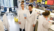


Dr Andre Neves
Position: Senior Research Associate
Personal home page:
http://www.researchgate.net/profile/Andre_Neves/
PubMed journal articles - click here
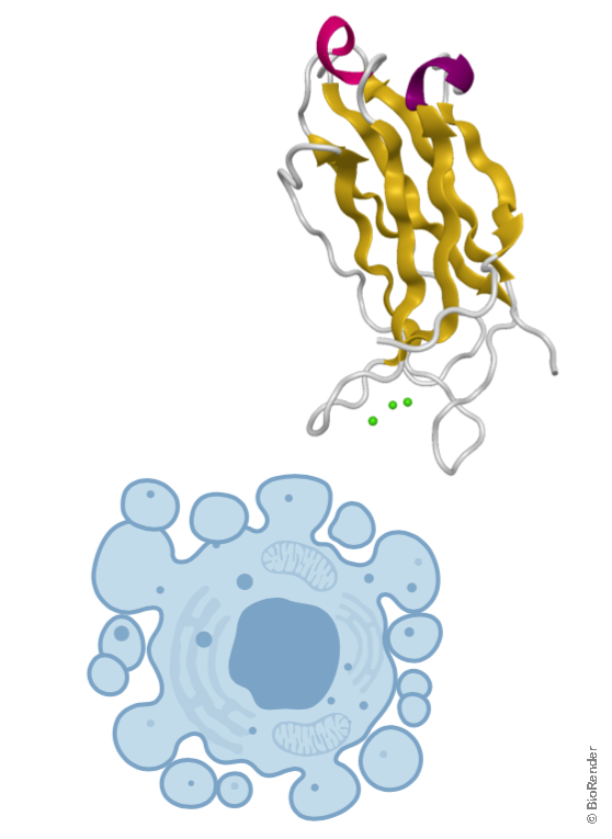
As a chemical engineer, I enjoy translating basic science into novel biomedical technologies. I am interested in the use of molecular imaging techniques for early cancer diagnosis and for better assessing response to cancer therapy. Currently, I'm planning a first-in-human clinical trial of a novel cell death imaging agent, C2Am. This new imaging scan will enable clinicians to assess more accurately and rapidly the patient's response to cancer therapies, potentially leading to better outcomes and significant cost savings.
Symplectic Elements feed provided by Research Information, University of Cambridge
[1] Rapid Imaging of Tumor Cell Death in vivo using the C2A domain of Synaptotagmin-I.
Neves AA, Xie B, Fawcett S, Alam IS, Witney TH, de Backer MM, Summers J, Hughes W, McGuire S, Soloviev D, Miller J, Howat WJ, Hu DE, Rodrigues TB, Lewis DY, Brindle KM.
J Nucl Med. 2017 58(6), 881-7.
[2] Imaging Glycosylation In Vivo by Metabolic Labeling and Magnetic Resonance Imaging.
Neves AA, Wainman YA, Wright A, Kettunen MI, Rodrigues TB, McGuire S, Hu DE, Bulat F, Geninatti Crich S, Stöckmann H, Leeper FJ, Brindle KM.
Angew Chem Int Ed Engl. 2016 Jan 22;55(4):1286-90.
[3] Molecular imaging using fluorescent lectins permits rapid endoscopic identification of dysplasia in Barrett's esophagus.
Bird-Lieberman EL, Neves AA, Lao-Sirieix P, O'Donovan M, Novelli M, Lovat LB, Eng WS, Mahal LK, Brindle KM & Fitzgerald RC
Nat Med. 2012 18(2):315-21.