


Professor Kevin Brindle
Position: Professor
Personal home page:
https://www.cruk.cam.ac.uk/research-groups/brindle-group
Email:
imaging@cancer.cam.ac.uk
PubMed journal articles - click here
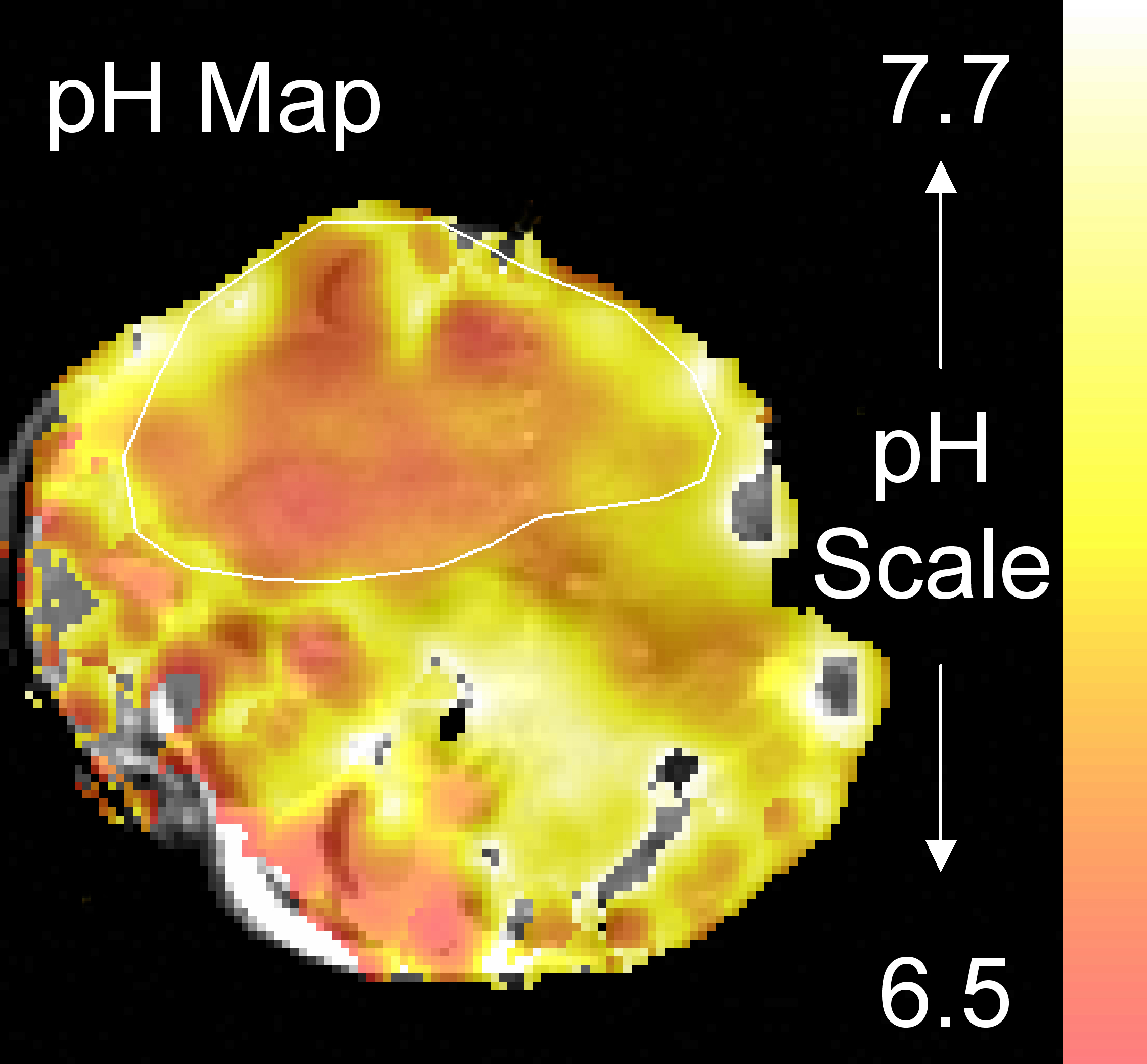
Magnetic resonance imaging (MRI) is a well-established, clinically applicable, tool for determining tissue morphology. The techniques of molecular imaging seek, through the use of appropriate probe molecules, to transfer into the MR image of tissue anatomy, information about underlying tissue biochemistry and physiology. We are developing novel magnetic resonance-based molecular imaging techniques to detect the early responses of tumours to therapy, with a view to translating these into clinical application. This has included methods for detecting and predicting responses to an anti-vascular drug and for detecting early tumour responses to immunotherapy. An early apoptotic response following treatment with a chemotherapeutic drug is a good prognostic indicator for treatment outcome. Therefore, a major focus is the development of magnetic resonance imaging (MRI) and spectroscopy (MRS) methods for the non-invasive detection of tumour cell death in vivo.
Symplectic Elements feed provided by Research Information, University of Cambridge
Day, S. E., Kettunen, M. I., Gallagher, F. A., Hu, D.-E., Lerche, M., Wolber, J., Golman, K., Ardenkjaer-Larsen, J. H., and Brindle, K. M. (2007) Detecting tumor response to treatment using hyperpolarized 13C magnetic resonance imaging and spectroscopy. Nature Medicine 13, 1382 ? 1387. Krishnan, A.S., Neves, A. A., de Backer, M. M., Hu, D.-E., Davletov, B., Kettunen, M. I., and Brindle, K. M. (2008) Detection of cell death in tumors using MRI and a gadolinium-based targeted contrast agent. Radiology 246, 854-862. Gallagher, F. A., Kettunen, M. I., Day, S. E., Hu, D.-E., Ardenkjær-Larsen, J. H., in ?t Zandt, R., Jensen, P. R., Karlsson, M., Golman, K., Lerche, M. H., and Brindle, K. M. (2008) Magnetic resonance imaging of pH in vivo using hyperpolarized 13C-labeled bicarbonate. Nature 453, 940-943.